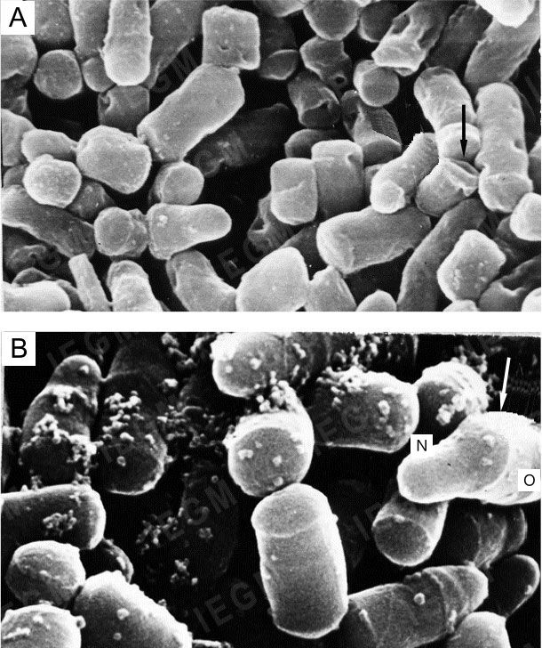
Scanning electron microscopy of Rhodococcus ruber cells grown on nutrient agar
Area near the top of a one-day colony
A – strain IEGM 565, x16,000. Peeling of the “old” peptidoglycan fragment from the cell surface (arrow); B – strain IEGM 333, x 30,000. Short rods. Ring-shaped protrusions (arrow) divide the cell into “new” (N) and “old” (O) growth zones