Emblem of IEGM Collection

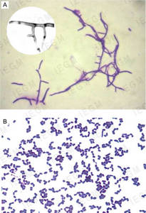
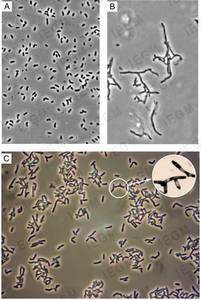
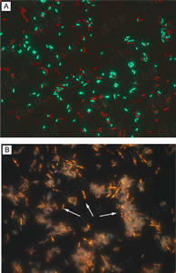
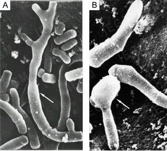
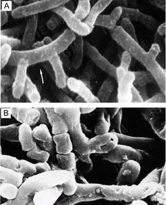
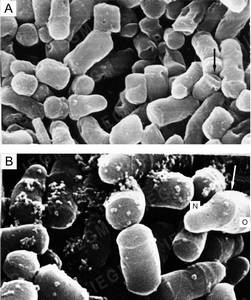
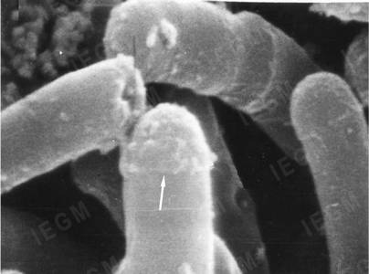
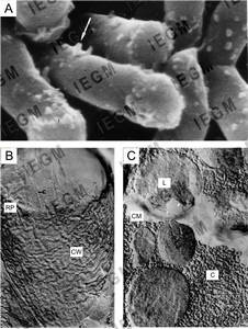
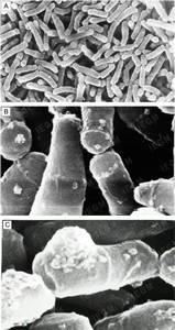
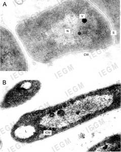
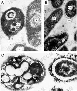
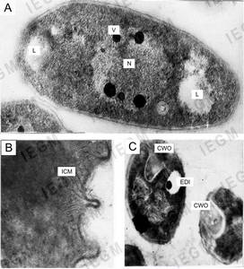
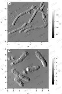
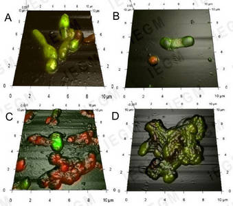
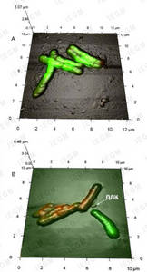
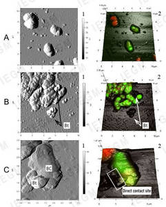
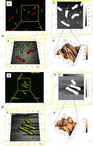
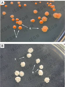
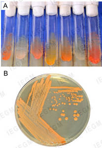
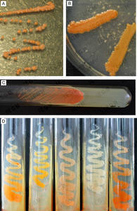
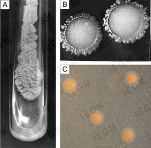
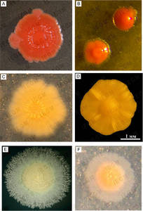
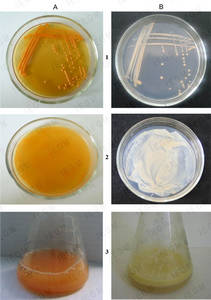
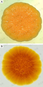
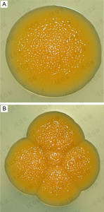
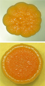
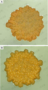
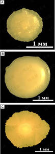
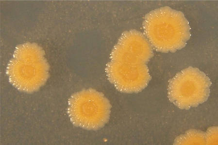
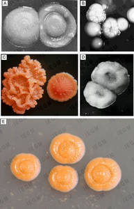
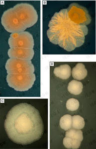
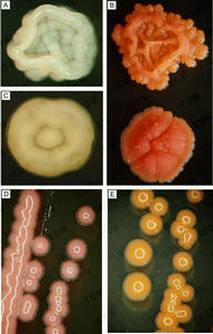
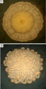
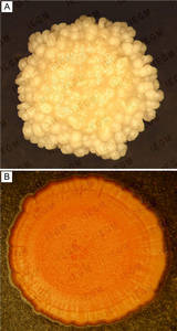
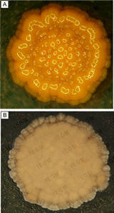
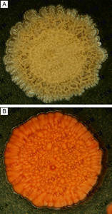
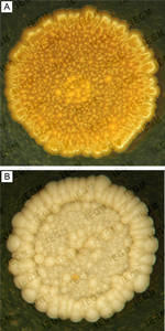
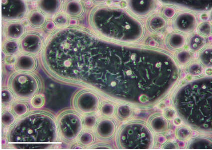
Gallery of the IEGM Collection Images


Stained smears from pure cultures. Light microscopy Cell morphology of R. ruber cells grown on nutrient agar. Phase contrast microscopy Pure culture preparations. Luminescence microscopy Scanning electron microscopy of R. ruber IEGM 333 cells grown on nutrient agar Scanning electron microscopy of R. ruber cells of the intermediate zone of a one-day colony grown on nutrient agar Scanning electron microscopy of R. ruber cells grown on nutrient agar Scanning electron microscopy of the snapping division of R. ruber IEGM 333 cells (magnification x 50,000) grown on nutrient agar Propane-grown R. ruber cells under scanning electron microscope (A) and their chips obtained by cryofractography (B, C) Scanning electron microscopy. R. ruber IEGM 87 cells grown on mineral salts agar in the presence of propane Ultra thin sections of R. ruber cells grown on nutrient agar Ultra thin sections of R. ruber IEGM 333 (A, C) and IEGM 565 (B) grown on MSM in the presence of propane (A, B) or n-hexadecane (C) Ultra thin sections of R. ruber cells grown on MSM with propane Atomic force microscopy (AFM). R. ruber IEGM 231 grown on nutrient agar Combined 3D AFM-CLSM images of R. ruber IEGM 346 grown in MSM for 10 days in the presence of glucose and an ecotoxicant Combined 3D AFM-CLSM image of immobilized R. erythropolis IEGM 267 cells after incubation with (B) or without (A) dehydroabietic acid AFM images (1) and combined 3D AFM-CLSM images (2) of R. rhodochrous IEGM 66 without (A) and with (B, C) betulin CLSM (a), AFM (b), and AFM-CLSM (c) images and an AFM image of the cell surface (d) of R. ruber IEGM 231 after 24-h incubation with (B) or without (A) acetone Dissociation of nutrient agar-grown Rhodococcus colonies into S- and R-forms Growth of rhodococci on nutrient agarMacromorphological characteristics of Rhodococcus members R. ruber IEGM 333 on mineral salts agar in the presence of propane R. ruber colonies on nutrient agar: (A) strain IEGM 333, (B) strain IEGM 71; on mineral salts agar in the presence of n-hexadecane: (C) strain IEGM 235, (D) strain IEGM 231 or propane: (E) strain IEGM 231, (F) strain IEGM 333 Comparative growth of R. ruber IEGM 235 on different nutrient media for five days Macrocolony of R. ruber IEGM 231 on nutrient agar Individual colony (A) and a macrocolony of 5 CFUs (B) of R. ruber IEGM 342 on nutrient agar Macrocolonies of R. ruber IEGM 231 on nutrient agar Rhodococcus macrocolonies with secondary growth, firmly attached to the surface of nutrient agar due to the formation of substrate mycelium: (A) R. aetherivorans IEGM 911T and (B) R. ruber IEGM 319 Morphological diversity of R. erythropolis IEGM 201 colonies depending on the carbon source used Colonies of R. ruber IEGM 236 on mineral salts agar with n-hexadecane S- and R-colonies of rhodococci on nutrient agar Colonies of R. aetherivorans IEGM 1250 (A, B) and R. opacus IEGM 261 (C, D) on mineral salts agar with n-hexadecane Colonies of different Rhodococcus species on nutrient agar Colonies of R. ruber IEGM 231 on mineral salts agar in the presence of propane (A), and colonies of R. pyridinivorans IEGM 1142 in the presence of n-hexadecane (B) Colonies of R. jostii IEGM 60 (A) and R. ruber IEGM 233 (B) Colonies of R. fascians IEGM 43 (A) and R. opacus IEGM 56 (B) Colonies of R. erythropolis IEGM 766 (A) and R. aetherivorans IEGM 1250 (B) Colonies of R. yunnanensis IEGM 1323 (A) and R. maanshanensis IEGM 1396 (B) R. ruber IEGM 235 cells inside a hydrocarbon drop (n-hexadecane)